Arterial Blood Gas (ABG) Interpretation: Complete Clinical Masterclass
Master step-by-step ABG interpretation, Henderson-Hasselbalch equations, Anion Gap, Delta-Delta ratio, Winter's formula, and real worked case vignettes.
Table of Contents (14 Sections)

Arterial Blood Gas (ABG) Interpretation: Complete Clinical Masterclass
AG = 12 ± 4 mEq/L (Na⁺ - (Cl⁻ + HCO₃⁻)). Correct for hypoalbuminemia (+2.5 mEq/L per 1 g/dL albumin drop below 4.0).
- Delta Ratio (Δ/Δ): (AG - 12) / (24 - HCO₃⁻): <0.8 = Mixed High AG + Normal AG Acidosis | 0.8–2.0 = Pure High AG Acidosis | >2.0 = High AG Acidosis + Pre-existing Metabolic Alkalosis.
- Oxygenation Status: P/F Ratio (PaO₂ / FiO₂): Normal >400 | Mild ARDS 200–300 | Moderate 100–200 | Severe <100 mmHg.---
📌 Interactive Table of Contents (Index)
- 1. Introduction & Clinical Indications
- 2. Pre-Analytical Errors & Sample Integrity
- 3. Normal ABG Reference Ranges & VBG Comparison
- 4. Universal 6-Step ABG Interpretation Algorithm
- 5. Acid-Base Disorders & Etiology Mnemonics
- 6. Advanced Equations: Winter's Formula & Delta Ratio
- 7. Flagship Worked Clinical Case Study
- 8. Oxygenation, A-a Gradient & P/F Ratio
- 9. High-Yield Clinical Pearls & Diagnostic Pitfalls
---
1. Introduction & Clinical Indications
Arterial blood gas (ABG) analysis is one of the most vital bedside diagnostic tests in emergency medicine, intensive care units (ICU), and acute inpatient wards. It provides rapid data regarding arterial oxygenation, alveolar ventilation, systemic acid-base equilibrium, and perfusion status.
Primary Clinical Indications
- Acute Respiratory Failure: Differentiate hypoxemic vs hypercapnic respiratory failure in asthma, COPD, pulmonary edema, or ARDS.
- Shock States & Hypoperfusion: Quantify metabolic acidosis and monitor serial lactate clearing in septic, cardiogenic, or hypovolemic shock.
- Altered Mental Status & Coma: Unmask acute hypercapnic encephalopathy (PaCO₂ > 70 mmHg) or toxic ingestions.
- Renal Failure & Electrolyte Emergencies: Evaluate severe metabolic acidosis in Acute Kidney Injury (AKI) or ESRD.
- Ventilator Titration: Guide minute ventilation adjustments, PEEP titration, and non-invasive ventilation (NIV/BiPAP).
---
2. Pre-Analytical Errors & Sample Integrity
More than 60% of blood gas errors occur prior to laboratory analysis. Sample integrity is paramount for clinical decision-making.
---
3. Normal ABG Reference Ranges & VBG Comparison
Understanding arterial reference values and how they compare to Venous Blood Gas (VBG) parameters saves patients from unnecessary arterial punctures.
| Parameter | Arterial Blood Gas (ABG) | Peripheral Venous (VBG) | Central Venous (CVBG) | Clinical Significance |
|---|---|---|---|---|
| pH | 7.35 – 7.45 | 0.03 – 0.05 lower | 0.03 – 0.05 lower | Systemic H⁺ ion concentration |
| PaCO₂ | 35 – 45 mmHg | 4.0 – 8.0 mmHg higher | 4.0 – 6.0 mmHg higher | Respiratory acid component |
| HCO₃⁻ | 22 – 26 mEq/L | 1.0 – 2.0 mEq/L higher | 1.0 – 2.0 mEq/L higher | Renal metabolic base component |
| PaO₂ | 80 – 100 mmHg | Unreliable for O₂ | Unreliable for O₂ | Arterial oxygen tension |
| SaO₂ | 95% – 100% | 70% – 75% | 70% – 75% (ScvO₂) | Hemoglobin oxygen saturation |
---
4. Universal 6-Step ABG Interpretation Algorithm
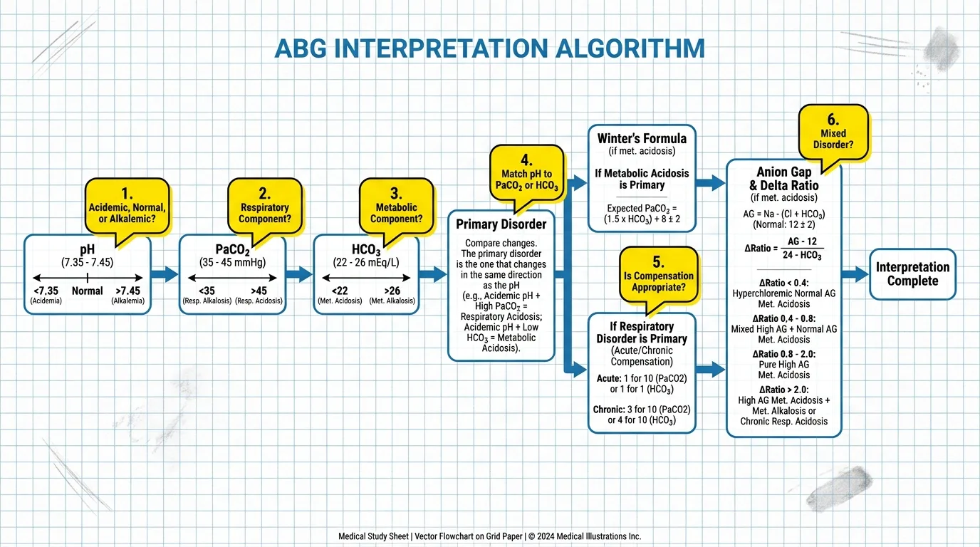
Follow this systematic step-by-step workflow on every blood gas report:
- Step 1: Check pH Status
- -
pH < 7.35: Acidemia - -
pH > 7.45: Alkalemia - -
pH 7.35 – 7.45: Normal or Fully Compensated / Mixed Disorder - Step 2: Check Respiratory Component (PaCO₂)
- -
PaCO₂ > 45 mmHg: Respiratory Acidosis (Hypoventilation) - -
PaCO₂ < 35 mmHg: Respiratory Alkalosis (Hyperventilation) - Step 3: Check Metabolic Component (HCO₃⁻)
- -
HCO₃⁻ < 22 mEq/L: Metabolic Acidosis - -
HCO₃⁻ > 26 mEq/L: Metabolic Alkalosis - Step 4: Determine Primary Disorder
- - Match the direction of pH with PaCO₂ or HCO₃⁻.
- Step 5: Calculate Expected Compensation
- - Determine if compensation is expected, incomplete, or represents a mixed disorder.
- Step 6: Calculate Anion Gap & Delta Ratio
- -
AG = Na⁺ - (Cl⁻ + HCO₃⁻)(Normal: 12 ± 4 mEq/L).
---
5. Acid-Base Disorders & Etiology Mnemonics
The four primary acid-base disturbances are categorized by mechanism and clinical presentation.
High Anion Gap Metabolic Acidosis (HAGMA) Mnemonic: MUDPILES
- M Methanol
- U Uremia (Renal Failure)
- D Diabetic Ketoacidosis (DKA) / Alcoholic Ketoacidosis
- P Propylene Glycol / Paraldehyde
- I Isoniazid / Iron
- L Lactic Acidosis (Type A: Hypoxia/Shock | Type B: Metformin/Malignancy)
- E Ethylene Glycol
- S Salicylates (Aspirin Toxicity)
Normal Anion Gap Metabolic Acidosis (NAGMA) Mnemonic: HARDCARD
- H Hyperalimentation (TPN)
- A Acetazolamide (Carbonic Anhydrase Inhibitors)
- R Renal Tubular Acidosis (RTA Types I, II, IV)
- D Diarrhea (Loss of Bicarbonate)
- C Uretero-sigmoidostomy
- A Addison's Disease
- R Resuscitation with Saline (Hyperchloremic Acidosis)
- D D-Lactic Acidosis
---
6. Advanced Equations: Winter's Formula & Delta Ratio
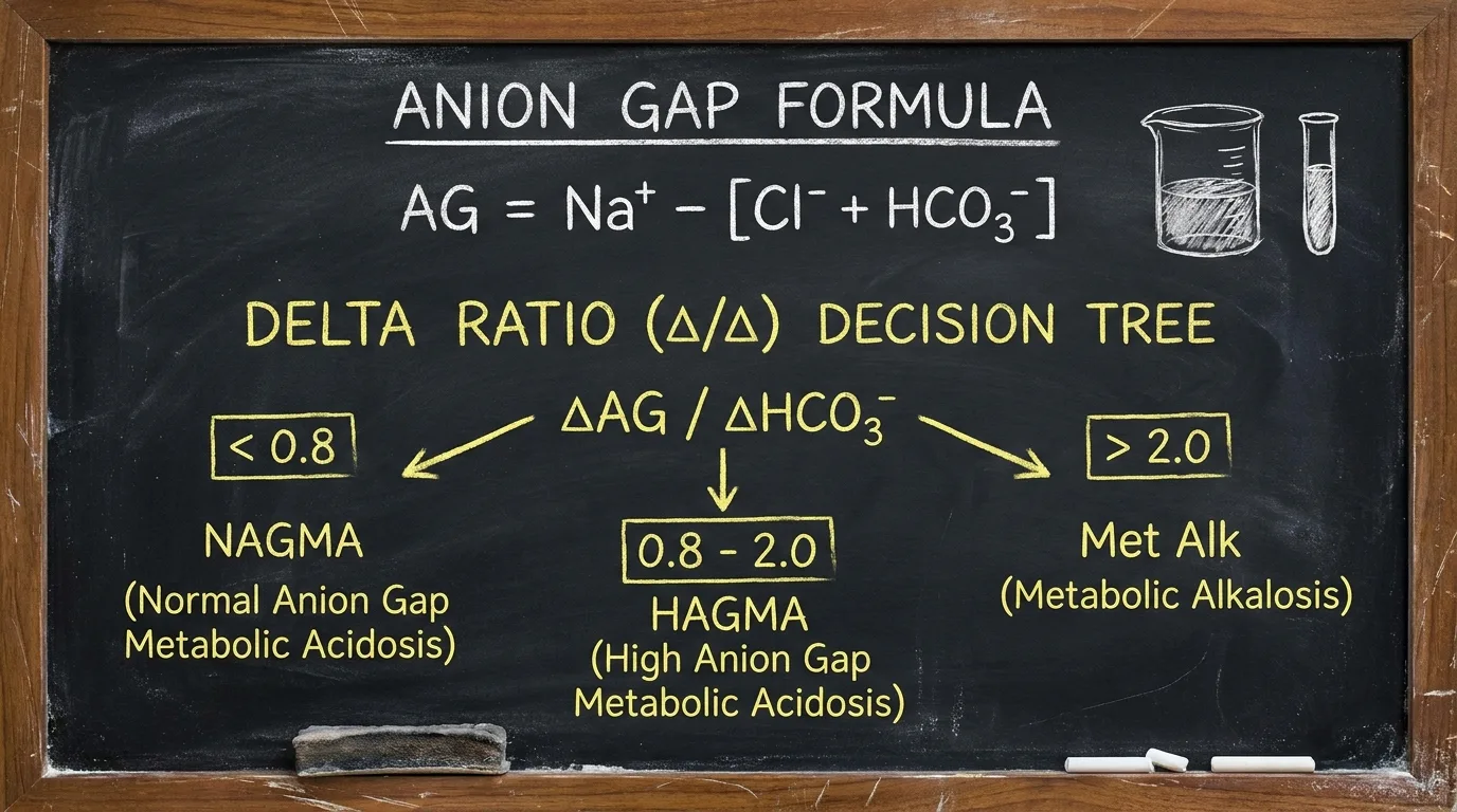
For complex ICU patients, standard interpretation is insufficient. Use these target formulas:
▶ Winter's Formula for Metabolic Acidosis Compensation
Calculates expected respiratory compensation (PaCO₂) in primary metabolic acidosis:
Expected PaCO₂ = (1.5 × HCO₃⁻) + 8 ± 2
- Actual PaCO₂ = Expected PaCO₂: Adequate respiratory compensation.
- Actual PaCO₂ > Expected PaCO₂: Concurrent Respiratory Acidosis (Respiratory failure / hypoventilation).
- Actual PaCO₂ < Expected PaCO₂: Concurrent Respiratory Alkalosis (Hyperventilation / Sepsis).
▶ Anion Gap Albumin Correction Formula
Hypoalbuminemia lowers measured anion gap. Correct AG to avoid missing hidden HAGMA:
Corrected AG = Measured AG + 2.5 × (4.0 - Measured Albumin in g/dL)
▶ Delta Ratio (Δ/Δ) for Mixed Metabolic Disorders
Determines if a second metabolic disorder coexists with High AG Metabolic Acidosis:
Delta Ratio = (Measured AG - 12) / (24 - Measured HCO₃⁻)
- < 0.8: Mixed High AG Acidosis + Normal AG Acidosis (e.g., DKA + Diarrhea).
- 0.8 – 2.0: Pure High Anion Gap Metabolic Acidosis (e.g., Uncomplicated DKA).
- > 2.0: High AG Acidosis + Pre-existing Metabolic Alkalosis (e.g., DKA + Severe Vomiting).
---
7. Flagship Worked Clinical Case Study
Worked Case: Diabetic Ketoacidosis (DKA) Analysis
Case StudyA 24-year-old patient with type 1 diabetes presents to the ED with lethargy, vomiting, and deep Kussmaul breathing.
#### 📊 Patient Lab & Vitals Panel
| Diagnostic Parameter | Measured Value | Normal Reference | Primary Interpretation |
|---|---|---|---|
| pH | 7.18 | 7.35 – 7.45 | Severe Acidemia |
| PaCO₂ | 20 mmHg | 35 – 45 mmHg | Hypocapnia (Hyperventilation) |
| HCO₃⁻ | 7 mEq/L | 22 – 26 mEq/L | Severe Metabolic Base Deficit |
| Sodium (Na⁺) | 138 mEq/L | 135 – 145 mEq/L | Serum Electrolytes |
| Chloride (Cl⁻) | 98 mEq/L | 95 – 105 mEq/L | Serum Electrolytes |
| Anion Gap (AG) | 33 mEq/L | 12 ± 4 mEq/L | High Anion Gap (+21 mEq/L) |
#### ⚡ 6-Step Clinical Decision Flowchart
Step 1: Systemic Status   →  pH 7.18 (< 7.35) = Severe Acidemia
Step 2: Respiratory Axis   →  PaCO₂ 20 mmHg (< 35) = Respiratory Alkalosis direction
Step 3: Metabolic Axis     →  HCO₃⁻ 7 mEq/L (< 22) = Metabolic Acidosis direction
Step 4: Primary Disorder   →  Metabolic Acidosis (pH matches HCO₃⁻ shift)
Step 5: Compensation Check →  Expected PaCO₂ = (1.5 × 7) + 8 ± 2 = 18.5 ± 2 mmHg
                             Actual PaCO₂ (20 mmHg) is within 16.5–20.5 range
                             Result: Appropriate Respiratory Compensation (Kussmaul breathing)
Step 6: Anion Gap & Delta  →  AG = 138 - (98 + 7) = 33 mEq/L (High AG Acidosis)
                             Delta Ratio = (33 - 12) / (24 - 7) = 21 / 17 = 1.23
                             Diagnosis: Pure High Anion Gap Metabolic Acidosis (DKA)
8. Oxygenation, A-a Gradient & P/F Ratio
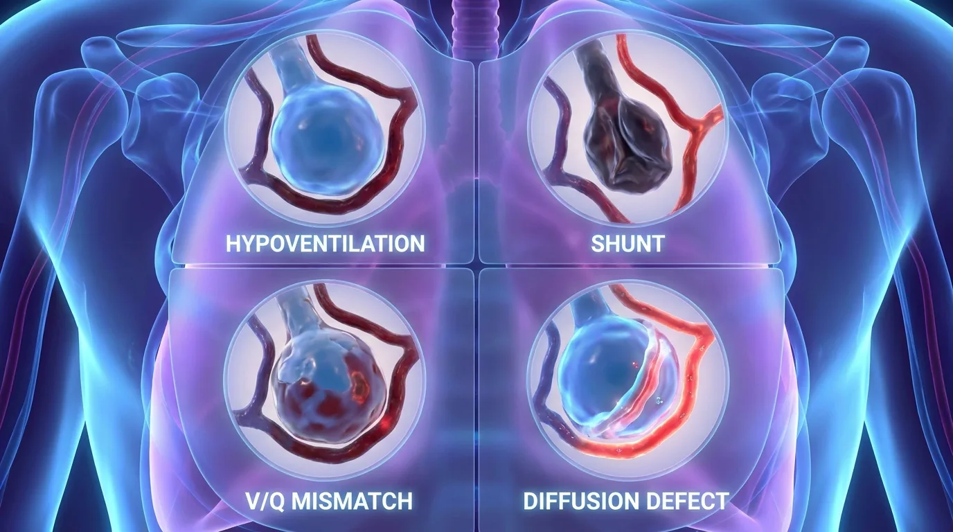
Evaluating tissue oxygenation is as critical as acid-base analysis.
- P/F Ratio (PaO₂ / FiO₂): Standard diagnostic criteria for ARDS (Berlin Definition).
- - Normal: >400 mmHg
- - Mild ARDS: 200 – 300 mmHg
- - Moderate ARDS: 100 – 200 mmHg
- - Severe ARDS: <100 mmHg
- Alveolar-Arterial (A-a) Gradient Formula:
P(A-a)O₂ = [FiO₂ × (Patm - PH₂O) - (PaCO₂ / 0.8)] - PaO₂- - Normal A-a Gradient:
< (Age / 4) + 4 - - Elevated A-a Gradient indicates V/Q mismatch, shunt, or diffusion impairment.
---
9. High-Yield Clinical Pearls & Diagnostic Pitfalls
Clinically Related Case Posters
Excessive Urination and Confusion
Diabetic Retinopathy. Peripheral Neuropathy. Undiagnosed Hypertension
Severe Shortness of Breath and Increased Cough in an Elderly Smoker
Cor Pulmonale (Right Heart Failure). Lung Cancer. Cachexia. Normocytic Anem...
Severe Breathlessness and Wheezing in a Young Student
Allergic Rhinitis. Eczema
Decreased Urine Output and Severe Weakness
Dehydration. Gastroenteritis. Undiagnosed Hypertension
Test Your Decision-Making in Acute Severe Asthma Exacerbation with Respiratory Acidosis
Experience this exact clinical scenario in the Bedside Rounds interactive simulator. Order investigations, manage door-to-needle timing, and witness real-time physiological feedback.