Acute Dyspnea Clinical Guide: Emergency Diagnosis & Management
Master acute breathlessness evaluation, BLUE protocol lung ultrasound, cardiac vs pulmonary dyspnea differentiation, and NIV airway management.
Table of Contents (13 Sections)
Acute Dyspnea Clinical Guide: Emergency Diagnosis & Management
≤ 15 minutes of acute hypercapnic respiratory failure arrival to reduce intubation rates by 65%.---
📌 Interactive Table of Contents (Index)
- 1. Introduction & Initial Resuscitation
- 2. Diagnostic Workup & Biomarkers
- 3. Cardiac vs Pulmonary Dyspnea Differentiation
- 4. BLUE Protocol Lung Ultrasound Algorithm
- 5. Non-Invasive Ventilation (NIV) & Airway Protocol
- 6. Advanced Respiratory Equations & Formulas
- 7. Flagship Worked Clinical Case Study
- 8. High-Yield Clinical Pearls & Diagnostic Traps
---
1. Introduction & Initial Resuscitation
Acute dyspnea (shortness of breath) is one of the most common and life-threatening chief complaints encountered in emergency medicine and intensive care.
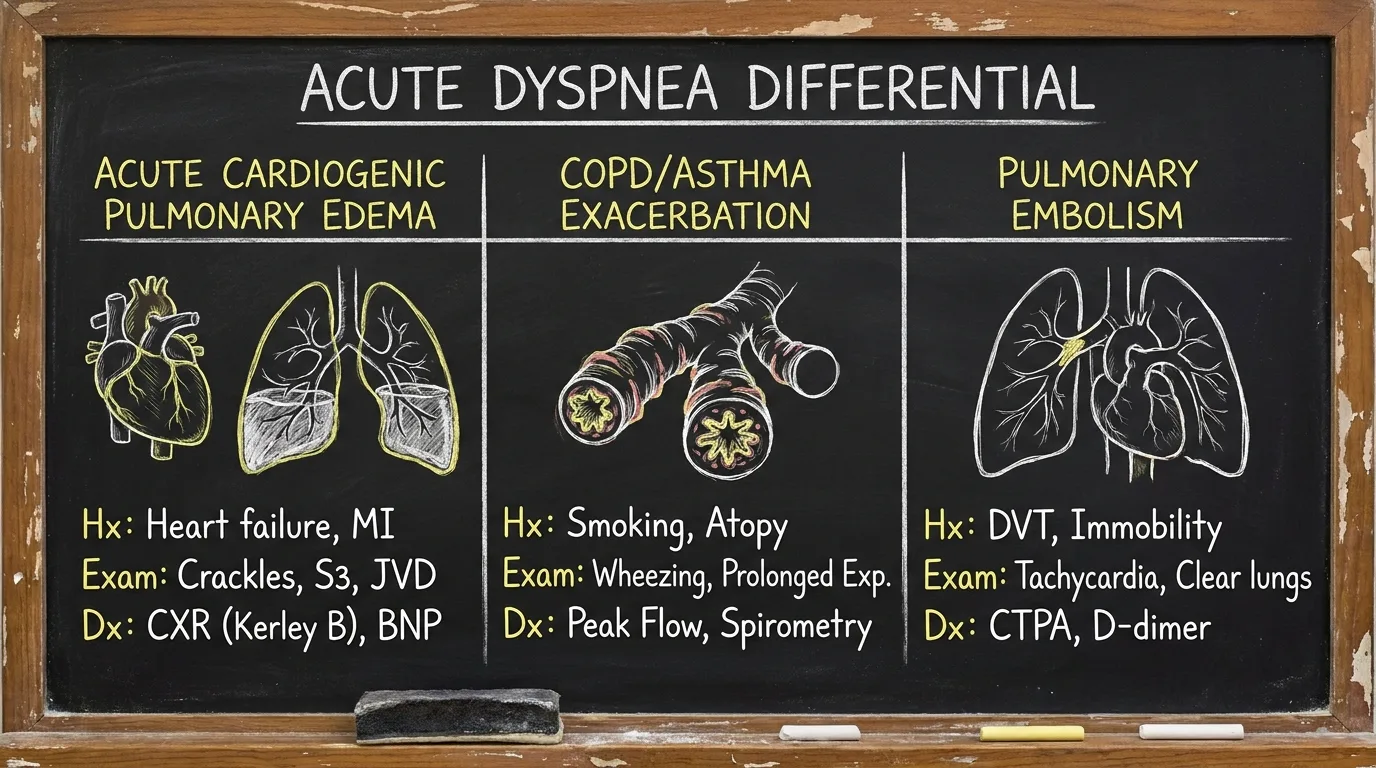
Initial ABCDE Triage Workflow
- Airway & Breathing: Assess stridor, accessory muscle use, and silent chest (impending respiratory arrest).
- Oxygenation Target: Maintain SpO₂ 94%–98% in non-COPD patients (88%–92% in chronic hypercapnic COPD).
- Hemodynamic Support: Check BP and perfusion. Flash pulmonary edema presents with hypertensive emergency (BP > 180/100 mmHg).
---
2. Diagnostic Workup & Biomarkers
Essential diagnostic modalities for rapid etiology identification:
| Test / Biomarker | Normal Reference Range | Diagnostic Interpretation in Acute Dyspnea | |
|---|---|---|---|
| NT-proBNP | < 300 pg/mL | > 450 pg/mL (<55 yr) | > 900 pg/mL (55–75 yr) = Acute Heart Failure |
| High-Sensitivity Troponin | < 14 ng/L | Elevated = Ischemic dyspnea / NSTEMI / Pulmonary Embolism | |
| D-Dimer | < 500 ng/mL FEU | High sensitivity to rule out PE (use YEARS / PERC criteria) | |
| Blood Gas (ABG) | pH 7.35–7.45 | PaCO₂ 35–45 | Detects acute respiratory acidosis, hypercapnia, and ARDS |
---
3. Cardiac vs Pulmonary Dyspnea Differentiation
Differentiating cardiogenic pulmonary edema from pulmonary airway obstruction (COPD/Asthma) is critical for acute therapy.
| Clinical Feature | Acute Cardiogenic Pulmonary Edema | COPD / Asthma Exacerbation |
|---|---|---|
| Primary Symptom | Orthopnea, Paroxysmal Nocturnal Dyspnea | Progressive exertional dyspnea, wheezing, cough |
| Physical Exam | Bilateral basalar crackles, S3 gallop, JVD | Expiratory wheezing, prolonged expiration, barrel chest |
| Lung POCUS | Bilateral symmetric B-lines (Lung Rockets) | A-lines with lung sliding, lung point if pneumothorax |
| Initial Medication | IV Furosemide + Sublingual Nitroglycerin + CPAP | Nebulized Albuterol/Ipratropium + IV Corticosteroids + BiPAP |
---
4. BLUE Protocol Lung Ultrasound Algorithm
Point-of-Care Ultrasound (POCUS) using the Bedside Lung Ultrasound in Emergency (BLUE) protocol provides >90% diagnostic accuracy within 3 minutes.
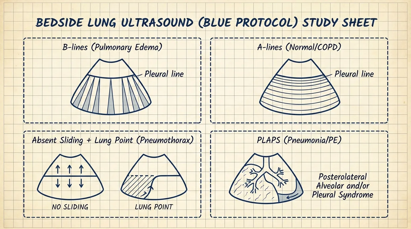
Key BLUE Protocol Profiles
- Profile B: Bilateral anterior B-lines with lung sliding → Acute Cardiogenic Pulmonary Edema.
- Profile A: Anterior A-lines with lung sliding + venous thrombosis on DVT scan → Pulmonary Embolism.
- Profile A (No DVT): Anterior A-lines + lung sliding → COPD / Asthma Exacerbation.
- Profile C: Anterior consolidation / focal B-lines → Pneumonia.
---
5. Non-Invasive Ventilation (NIV) & Airway Protocol
Initiate NIV promptly to improve work of breathing and reduce endotracheal intubation.
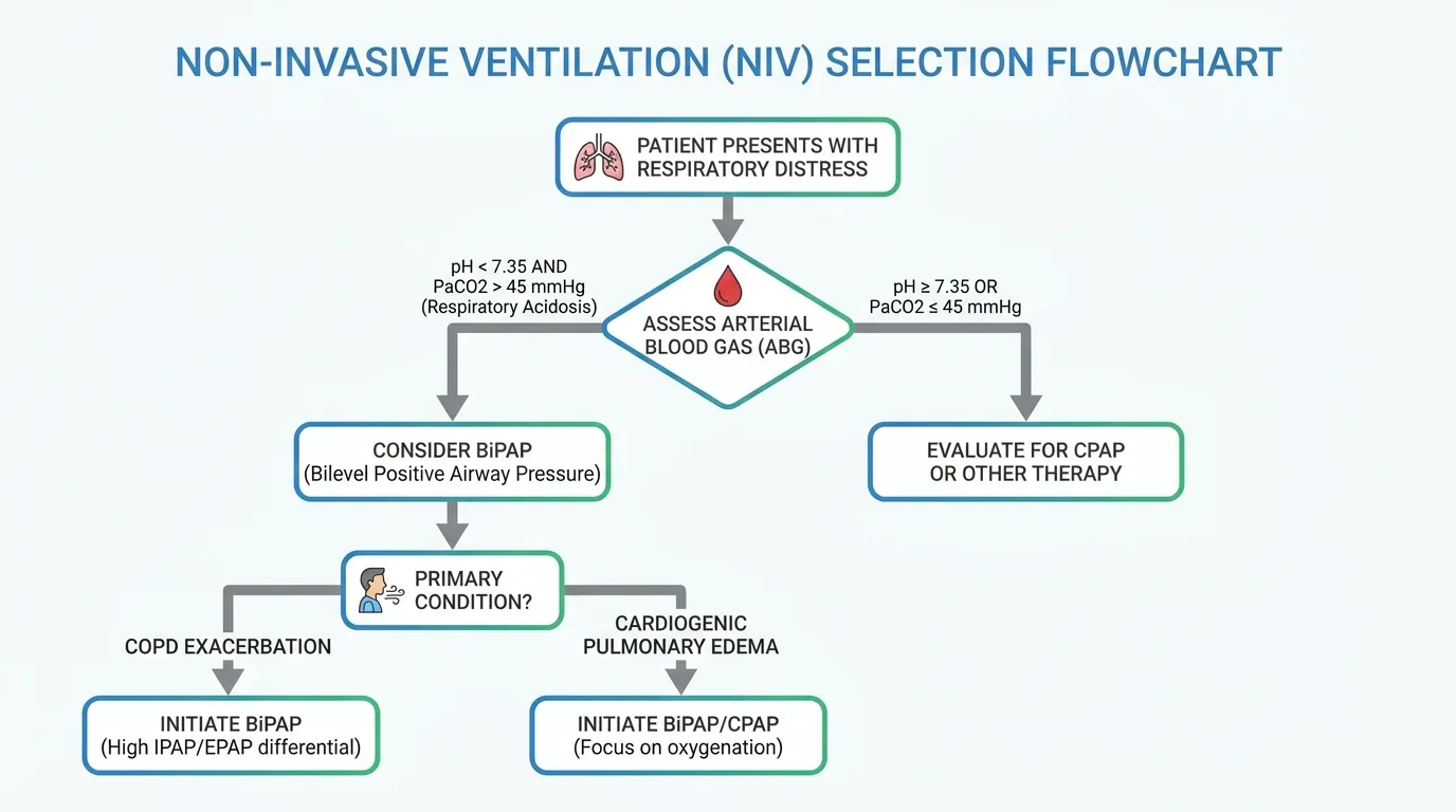
Mode Selection
- CPAP (Continuous Positive Airway Pressure): Preferred for acute cardiogenic pulmonary edema (5–10 cmH₂O).
- BiPAP (Bilevel Positive Airway Pressure): Preferred for acute hypercapnic COPD exacerbations (IPAP 10–12 / EPAP 4–5 cmH₂O).
---
6. Advanced Respiratory Equations & Formulas
▶ Rapid Shallow Breathing Index (RSBI) for Extubation Readiness
Formula: RSBI = Respiratory Rate (breaths/min) / Tidal Volume (Liters)
- RSBI < 105: High likelihood of successful extubation / weaning.
- RSBI > 105: High risk of weaning failure and re-intubation.
▶ P/F Ratio (PaO₂ / FiO₂) for ARDS Severity
- > 400 mmHg: Normal oxygenation.
- 200 – 300 mmHg: Mild ARDS.
- 100 – 200 mmHg: Moderate ARDS.
- < 100 mmHg: Severe ARDS.
---
7. Flagship Worked Clinical Case Study
Worked Case: Flash Pulmonary Edema in Hypertensive Crisis
Case StudyA 68-year-old female presents with severe respiratory distress, diaphoresis, and blood pressure of 210/115 mmHg.
#### 📊 Patient Lab & Vitals Panel
| Diagnostic Parameter | Measured Value | Reference Range | Diagnostic Interpretation |
|---|---|---|---|
| Blood Pressure | 210 / 115 mmHg | 120/80 mmHg | Hypertensive Emergency / Flash Edema |
| Heart / Resp Rate | 118 bpm / 34 bpm | 60–100 / 12–20 | Severe Tachypnea & Sympathetic Surge |
| SpO₂ (Room Air) | 78% | 95% – 100% | Severe Hypoxemic Failure |
| Lung POCUS | Bilateral B-lines | A-lines | Acute Pulmonary Edema |
| NT-proBNP | 4,200 pg/mL | <300 pg/mL | Markedly Elevated |
#### ⚡ Step-by-Step Clinical Decision Flowchart
Step 1: Triage Assessment  → SpO₂ 78% + BP 210/115 mmHg + Bilateral crackles = Flash Cardiogenic Pulmonary Edema
Step 2: Airway & Breathing  → Start CPAP 10 cmH₂O immediately + High-flow O₂ (Reduces preload & afterload)
Step 3: Afterload Reduction → High-dose Sublingual Nitroglycerin 0.8–1.2 mg q5min or IV Nitroglycerin infusion
Step 4: Diuresis             → IV Furosemide 40–80 mg IV bolus (once afterload reduction initiated)
Step 5: Outcome             → BP drops to 145/88 mmHg, SpO₂ improves to 96% on CPAP within 25 minutes
---
8. High-Yield Clinical Pearls & Diagnostic Traps
Clinically Related Case Posters
Progressive Breathlessness and Leg Swelling
Atrial Fibrillation. Chronic Obstructive Pulmonary Disease (COPD). Sleep Ap...
Sudden Shortness of Breath and Chest Pain
None
Severe Shortness of Breath and Increased Cough in an Elderly Smoker
Cor Pulmonale (Right Heart Failure). Lung Cancer. Cachexia. Normocytic Anem...
Acute Fever with Productive Cough
Type 2 Diabetes Mellitus. COPD. Chronic Kidney Disease. Benign Prostatic Hy...
Test Your Decision-Making in Acute Severe Dyspnea in Flash Pulmonary Edema
Experience this exact clinical scenario in the Bedside Rounds interactive simulator. Order investigations, manage door-to-needle timing, and witness real-time physiological feedback.