Acute STEMI Emergency Management: Step-by-Step Clinical Guide
Master acute STEMI diagnosis, 12-lead ECG coronary mapping, Sgarbossa criteria, primary PCI time targets, fibrinolysis, and shock management.
Table of Contents (11 Sections)
Acute STEMI Emergency Management: Step-by-Step Clinical Guide
≤ 10 minutes.
- Reperfusion Benchmarks: Primary PCI Door-to-Balloon target ≤ 60 minutes (or ≤ 120 minutes FMC-to-wire for hospital transfers).
- Fibrinolysis Protocol: Door-to-Needle target ≤ 30 minutes when Primary PCI delay exceeds 120 minutes.
- RV Infarct Rule: Avoid Nitrates, Morphine, and Diuretics in Right Ventricular Infarction (V4R STE); treat hypotension with IV Normal Saline boluses.---
📌 Interactive Table of Contents (Index)
- 1. Introduction & Pathophysiology
- 2. Emergency Triage & Diagnostic ECG Criteria
- 3. Coronary Territory Localization Table
- 4. Universal Step-by-Step STEMI Protocol
- 5. Reperfusion Therapy: Primary PCI vs Fibrinolysis
- 6. High-Risk STEMI Equivalents & Sgarbossa
- 7. Flagship Worked Clinical Case Study
- 8. High-Yield Clinical Pearls & Pitfalls
---
1. Introduction & Pathophysiology
Acute ST-Elevation Myocardial Infarction (STEMI) occurs due to acute transmural myocardial ischemia caused by thrombotic occlusion of an epicardial coronary artery following atherosclerotic plaque rupture or erosion.
Emergency Objectives
- Immediate ECG Acquisition: Obtain and interpret 12-lead ECG within 10 minutes of arrival.
- Rapid Reperfusion: Restore coronary blood flow via Primary Percutaneous Coronary Intervention (PCI) or Fibrinolysis.
- Prevent Complications: Prevent lethal ventricular fibrillation, cardiogenic shock, and acute papillary muscle rupture.
---
2. Emergency Triage & Diagnostic ECG Criteria
Diagnostic ST elevation is measured at the J-point in two anatomically contiguous leads:
| Lead Grouping | ST Elevation Threshold | Anatomical Lead Contiguity | High-Risk Features | ||
|---|---|---|---|---|---|
| Precordial V2–V3 | ≥ 2.5 mm (men <40) | ≥ 2.0 mm (men ≥40) | ≥ 1.5 mm (women) | V1–V2, V2–V3, V3–V4 | Proximal LAD occlusion |
| All Other Leads | ≥ 1.0 mm in all other leads (I, aVL, II, III, aVF, V5, V6) | II/III/aVF, I/aVL, V5/V6 | Reciprocal ST depression | ||
| Right Leads (V4R) | ≥ 0.5 mm in lead V4R (in inferior MI) | V3R, V4R | RV infarction, preload dependence | ||
| Posterior (V7–V9) | ≥ 0.5 mm in leads V7, V8, V9 | V7–V9 | Reciprocal ST dep in V1–V3 |
---
3. Coronary Territory Localization Table
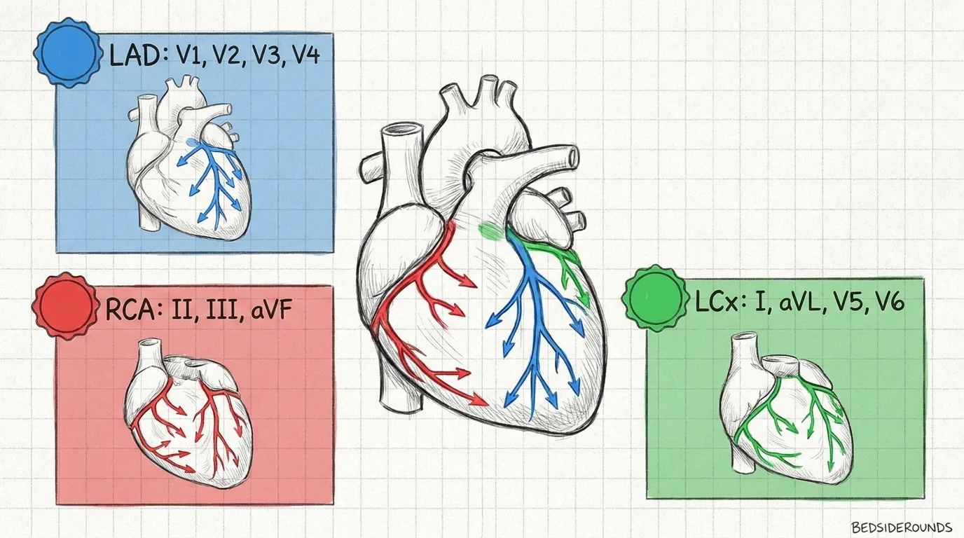
Identify culprit coronary arteries and reciprocal ECG changes:
| Infarct Territory | Primary ECG Leads | Culprit Coronary Artery | Reciprocal ST Changes |
|---|---|---|---|
| Anterior / Septal | V1, V2, V3, V4 | Left Anterior Descending (LAD) | Inferior (II, III, aVF) |
| Inferior | II, III, aVF | Right Coronary Artery (RCA 85%) | High Lateral (I, aVL) |
| Lateral | I, aVL, V5, V6 | Left Circumflex (LCx) / Diagonal | Inferior (II, III, aVF) |
| Right Ventricular | V3R, V4R | Proximal RCA | High Lateral |
---
4. Universal Step-by-Step STEMI Protocol
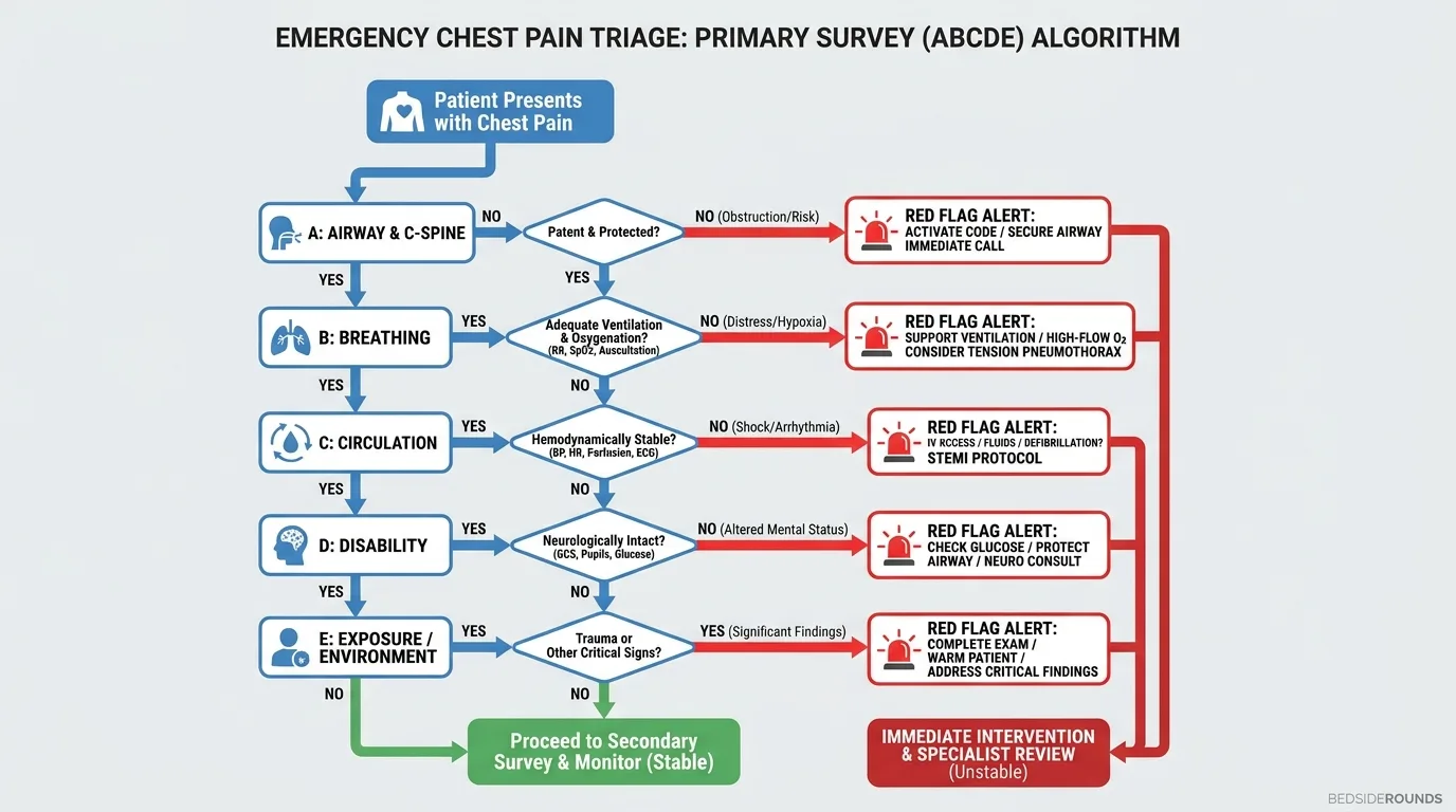
Follow this immediate resuscitation and antiplatelet regimen:
- Step 1: Emergency Triage & Oxygenation
- - Obtain 12-lead ECG within 10 minutes.
- - Give O₂ ONLY if SaO₂ < 90% (Hyperoxia causes coronary vasoconstriction!).
- Step 2: Dual Antiplatelet Therapy (DAPT)
- - Aspirin: 325 mg chewed immediately (non-enteric coated).
- - P2Y12 Inhibitor: Loading dose: Ticagrelor 180 mg OR Prasugrel 60 mg OR Clopidogrel 600 mg.
- Step 3: Anticoagulation & Anti-Ischemic Support
- - Unfractionated Heparin (UFH): 60 units/kg IV bolus (max 4000 U).
- - Nitroglycerin: Sublingual 0.4 mg every 5 min × 3 (Contraindicated if RV MI or PDE-5 inhibitor use!).
- Step 4: Reperfusion Activation
- - Activate Cath Lab immediately for Primary PCI.
---
5. Reperfusion Therapy: Primary PCI vs Fibrinolysis
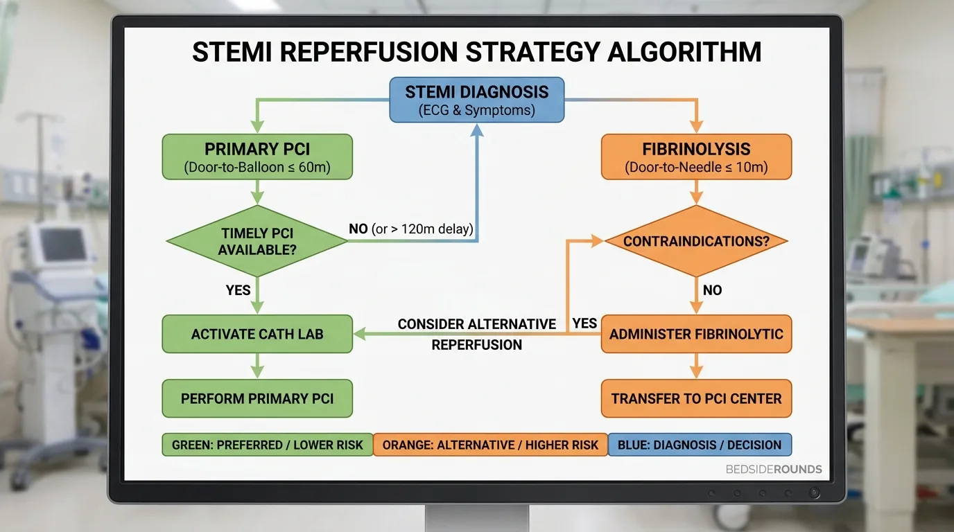
Choose the optimal reperfusion modality based on door-to-balloon time benchmarks:
| Reperfusion Modality | Target Time Benchmark | Primary Indications | Key Contraindications |
|---|---|---|---|
| Primary PCI | Door-to-Balloon ≤ 60 min (or ≤ 120 min for transfer) | Preferred for all STEMI | None (Life-saving emergency) |
| Fibrinolysis | Door-to-Needle ≤ 30 min | Only if PCI delay >120 min | Active bleeding, prior ICH, head trauma <3 mo |
---
6. High-Risk STEMI Equivalents & Sgarbossa
Recognize occlusion MI equivalents that require immediate Cath Lab activation despite absence of classic STE.
▶ De Winter Pattern (Proximal LAD Occlusion)
- 1–3 mm upsloping ST depression at J-point in V1–V6.
- Tall, prominent, symmetrical T waves in precordial leads.
- Represents 100% proximal LAD occlusion. Activate Cath Lab immediately!
▶ Smith-Modified Sgarbossa Criteria (STEMI in LBBB)
- 1.Concordant STE ≥ 1 mm in any lead with positive QRS (Score 5).
- 2.Concordant ST depression ≥ 1 mm in V1–V3 (Score 3).
- 3.Proportional Discordant STE:
(ST / S) ratio ≤ -0.25(Smith modification). - 4.
---
7. Flagship Worked Clinical Case Study
Worked Case: Inferior STEMI with Right Ventricular Infarction
Case StudyA 61-year-old male presents with severe pressure-like retrosternal pain, diaphoresis, and blood pressure of 82/50 mmHg.
#### 📊 Patient Lab & Vitals Panel
| Diagnostic Parameter | Measured Value | Reference Range | Diagnostic Interpretation |
|---|---|---|---|
| Blood Pressure | 82 / 50 mmHg | 120/80 mmHg | Hypotension / Preload Dependent |
| ECG II, III, aVF | 3 mm STE in III > II | Isoelectric | Acute Inferior STEMI (RCA) |
| ECG Lead V4R | 1.5 mm STE in V4R | Isoelectric | Right Ventricular (RV) Infarction |
| Reciprocal I, aVL | 2 mm ST depression | Isoelectric | High Lateral Reciprocal Changes |
| Troponin I | 14.2 ng/mL | <0.04 ng/mL | Acute Myocardial Necrosis |
#### ⚡ Step-by-Step Clinical Decision Flowchart
Step 1: ECG Recognition  → 3 mm ST elevation in inferior leads (III > II) + ST depression in aVL
Step 2: RV Lead Check    → Lead V4R shows 1.5 mm ST elevation = Acute RV Infarction confirmed
Step 3: Hemodynamic Rule → Hypotension (82/50 mmHg) due to RV failure & loss of preload
Step 4: Medication Trap  → CONTRAINDICATED: Nitrates, Morphine, & Diuretics (will cause fatal collapse!)
Step 5: Acute Therapy    → 1. Chewed Aspirin 325 mg + Ticagrelor 180 mg
                           2. IV Normal Saline 1000 mL bolus (Restores RV preload & BP)
                           3. Immediate Cath Lab activation (Door-to-Balloon Target ≤ 60 min)
---
8. High-Yield Clinical Pearls & Pitfalls
Clinically Related Case Posters
Crushing Chest Pain with Profuse Sweating
Type 2 Diabetes (poorly controlled). Essential Hypertension (on treatment)
Stable Angina with Multiple Cardiovascular Risk Factors
Type 2 Diabetes Mellitus. Hypertension. Dyslipidemia. Bronchial Asthma. Pep...
Persistent Fever and Breathlessness in a Young Man
Chronic Hepatitis C (untreated, diagnosed 2 years ago). Active intravenous ...
Test Your Decision-Making in Acute Inferior STEMI with Cardiogenic Shock
Experience this exact clinical scenario in the Bedside Rounds interactive simulator. Order investigations, manage door-to-needle timing, and witness real-time physiological feedback.