Chest Pain Emergency Evaluation: Step-by-Step Clinical Guide
Master acute chest pain risk stratification, HEART score calculation, 6 life-threatening etiologies, and emergency diagnostic algorithms.
Table of Contents (10 Sections)
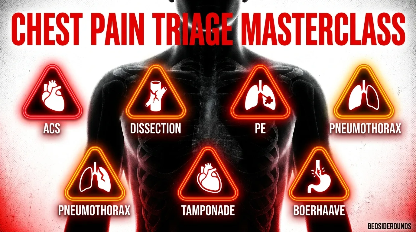
Chest Pain Emergency Evaluation: Step-by-Step Clinical Guide
≤ 10 minutes of ED arrival.
- HEART Score Rule: Score 0–3 = Low risk (discharge with outpatient follow-up) | Score 4–6 = Moderate risk (observe/admit) | Score 7–10 = High risk (early invasive cath).
- Aortic Dissection Red Flag: Sudden tearing pain radiating to interscapular back + BP differential > 20 mmHg between arms.---
📌 Interactive Table of Contents (Index)
- 1. Introduction & Initial Resuscitation
- 2. The 6 Life-Threatening Etiologies
- 3. HEART Score Risk Stratification Table
- 4. Universal Step-by-Step Triage Algorithm
- 5. Differential Diagnosis & Biomarker Comparison
- 6. Advanced Diagnostic Formulas
- 7. Flagship Worked Clinical Case Study
- 8. High-Yield Clinical Pearls & Pitfalls
---
1. Introduction & Initial Resuscitation
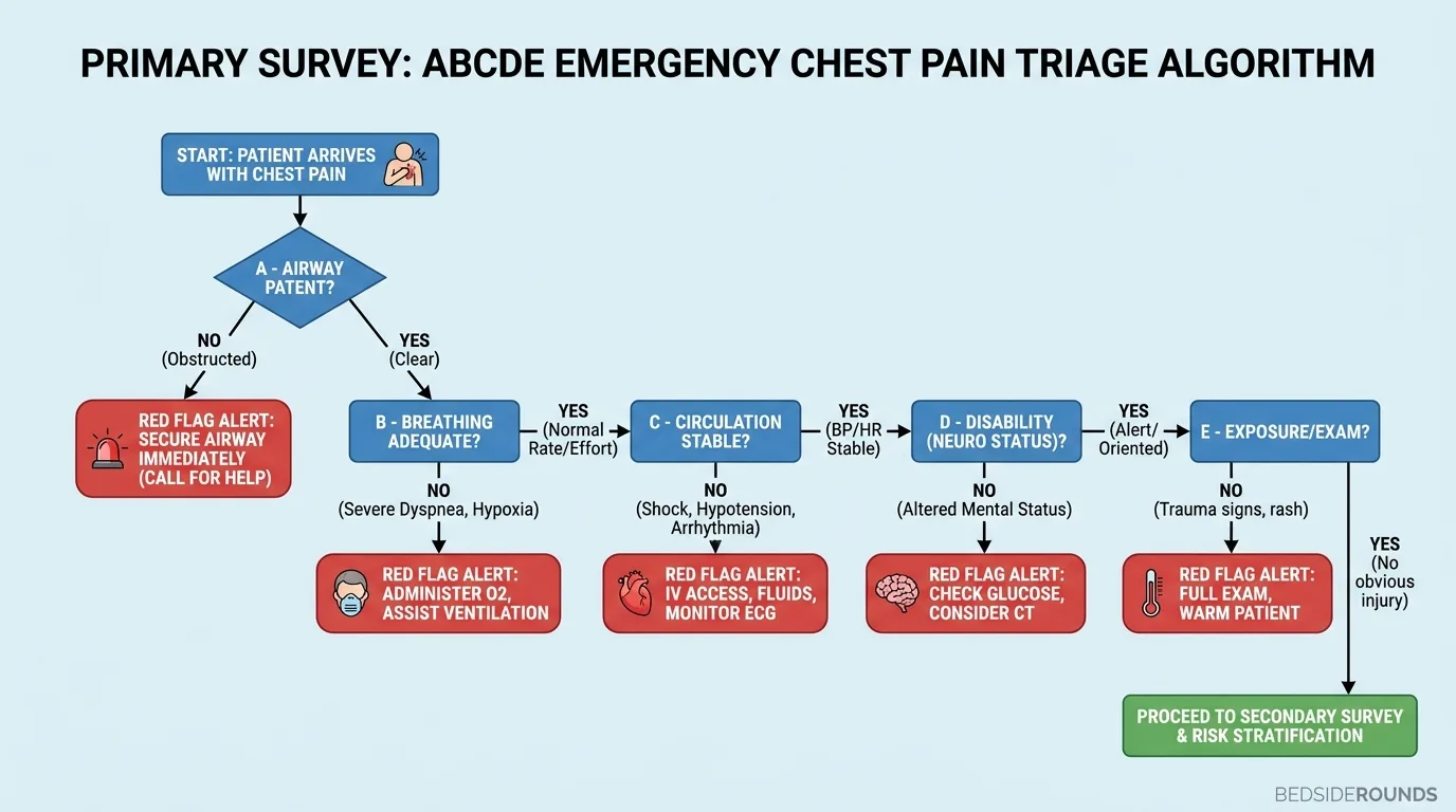
Acute chest pain is one of the most critical presentation complaints requiring rapid risk stratification to differentiate benign musculoskeletal pain from immediate lethal vascular emergencies.
---
2. The 6 Life-Threatening Etiologies
Systematically rule out these 6 killer etiologies during initial ED assessment:
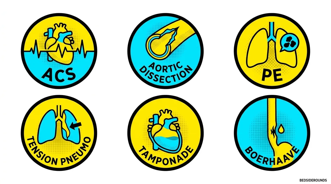
| Lethal Condition | Pathognomonic History / Exam | Diagnostic Gold Standard | Immediate Management |
|---|---|---|---|
| Acute Coronary Syndrome | Retrosternal pressure, radiation to left arm/jaw, diaphoresis | 12-Lead ECG + High-Sensitivity Troponin | Aspirin + DAPT + Cath Lab activation |
| Aortic Dissection | Sudden tearing pain to back, BP arm differential >20 mmHg | CT Angiography of Chest/Abdomen | IV Esmolol (Target HR <60, SBP <120) |
| Pulmonary Embolism | Pleuritic pain, acute dyspnea, tachycardia, DVT signs | CT Pulmonary Angiogram (CTPA) | Anticoagulation (Heparin) / Thrombolysis |
| Tension Pneumothorax | Absent breath sounds, tracheal deviation, hypotension | Bedside POCUS / Chest X-Ray | Needle decompression (2nd/5th ICS) |
| Cardiac Tamponade | Beck's Triad (Hypotension, JVD, muffled heart sounds) | Bedside Echo (POCUS) | Pericardiocentesis |
| Esophageal Rupture | Post-emesis chest pain, subcutaneous emphysema | CT Chest with Oral Contrast | IV Antibiotics + Surgical repair |
---
3. HEART Score Risk Stratification Table
The HEART score predicts 6-week Major Adverse Cardiac Events (MACE):
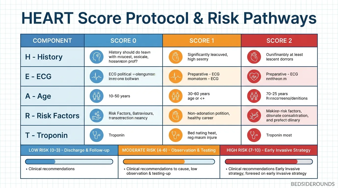
| HEART Category | Score 0 | Score 1 | Score 2 |
|---|---|---|---|
| H - History | Slightly suspicious | Moderately suspicious | Highly suspicious |
| E - ECG | Normal | Nonspecific repolarization | ST depression or elevation |
| A - Age | < 45 years | 45 – 64 years | ≥ 65 years |
| R - Risk Factors | 0 risk factors | 1 – 2 risk factors | ≥ 3 risk factors or atherosclerotic disease |
| T - Troponin | ≤ Normal limit | 1 – 3× Normal limit | > 3× Normal limit |
---
4. Universal Step-by-Step Triage Algorithm
- Step 1: 10-Minute ECG Check
- - Obtain 12-lead ECG immediately. If ST elevation present → Activate Cath Lab.
- Step 2: Check Vital Signs & Bilateral BPs
- - Measure blood pressure in both arms. A >20 mmHg difference strongly suggests Aortic Dissection.
- Step 3: Calculate HEART Score
- - Score 0–3: Low MACE risk (<1.7%). Safe for early discharge with outpatient stress testing.
- - Score 4–6: Moderate MACE risk (12–16%). Admit for serial troponins and inpatient evaluation.
- - Score ≥ 7: High MACE risk (>50%). Urgent Cardiology consultation & invasive angiography.
---
5. Differential Diagnosis & Biomarker Comparison
| Biomarker | Sensitivity / Specificity | Peak Time | Primary Clinical Role |
|---|---|---|---|
| High-Sensitivity Troponin I/T | >95% Sensitivity for ACS | 3–6 hours | Rule in / rule out myocardial necrosis |
| D-Dimer | High Sensitivity, Low Specificity | Immediate | Exclude PE & Dissection when low probability |
| CK-MB | Moderate Sensitivity | 12–24 hours | Re-infarction detection |
---
6. Advanced Diagnostic Formulas
▶ PERC Rule for Pulmonary Embolism Exclusion
If ALL 8 criteria are negative in a low-probability patient, PE is ruled out without D-dimer testing: 1. Age < 50 years 2. Heart Rate < 100 bpm 3. SpO₂ ≥ 95% on room air 4. No prior DVT/PE history 5. No recent surgery/trauma (< 4 weeks) 6. No hemoptysis 7. No estrogen use 8. No unilateral leg swelling
---
7. Flagship Worked Clinical Case Study
Worked Case: Acute Aortic Dissection Mimicking STEMI
Case StudyA 58-year-old male presents with sudden tearing chest pain radiating to his shoulder blades and BP of 190/110 mmHg in the right arm vs 142/80 mmHg in the left arm.
#### 📊 Patient Lab & Vitals Panel
| Diagnostic Parameter | Measured Value | Reference Range | Diagnostic Interpretation |
|---|---|---|---|
| Right Arm BP | 190 / 110 mmHg | 120/80 mmHg | Severe Hypertensive Crisis |
| Left Arm BP | 142 / 80 mmHg | 120/80 mmHg | 48 mmHg Arm Differential! |
| 12-Lead ECG | Sinus Tachy, Inferior ST dep | Isoelectric | Nonspecific Ischemia |
| CT Angiogram Chest | Type A Intimal Tear | Normal | Stanford Type A Aortic Dissection |
| Hs-Troponin | 0.08 ng/mL | <0.04 ng/mL | Mild secondary elevation |
#### ⚡ Step-by-Step Clinical Decision Flowchart
Step 1: Symptom Recognition  → Sudden tearing back pain + 48 mmHg BP differential between arms
Step 2: Medication Trap       → DO NOT give Aspirin, Heparin, or Cath Lab DAPT! (Fatal in Dissection!)
Step 3: Diagnostic Action     → Stat CT Angiography of Chest/Abdomen/Pelvis
Step 4: Acute HR & BP Control → Start IV Esmolol infusion (Target HR < 60 bpm, SBP < 120 mmHg)
Step 5: Surgical Action       → Immediate Cardiothoracic Surgery consultation for Type A Repair
---
8. High-Yield Clinical Pearls & Pitfalls
Clinically Related Case Posters
Crushing Chest Pain with Profuse Sweating
Type 2 Diabetes (poorly controlled). Essential Hypertension (on treatment)
Sharp Chest Pain Worsening when Lying Flat
Recent Upper Respiratory Tract Infection
Sudden Shortness of Breath and Chest Pain
None
Stable Angina with Multiple Cardiovascular Risk Factors
Type 2 Diabetes Mellitus. Hypertension. Dyslipidemia. Bronchial Asthma. Pep...
Test Your Decision-Making in Aortic Dissection Mimicking Acute MI
Experience this exact clinical scenario in the Bedside Rounds interactive simulator. Order investigations, manage door-to-needle timing, and witness real-time physiological feedback.