Syncope Risk Stratification: Step-by-Step Clinical Guide
Master syncope risk stratification, San Francisco Syncope Rule, Canadian Syncope Risk Score, cardiac vs vasovagal causes, and red flag ECG features.
Table of Contents (10 Sections)
Syncope Risk Stratification: Step-by-Step Clinical Guide
---
📌 Interactive Table of Contents (Index)
- 1. Introduction & Etiology Classification
- 2. Diagnostic Workup & ECG Red Flags
- 3. Cardiac vs Reflex Syncope Differentiation
- 4. San Francisco & Canadian Syncope Risk Scores
- 5. Structural & Channelopathy Red Flags Table
- 6. Advanced Diagnostic Equations
- 7. Flagship Worked Clinical Case Study
- 8. High-Yield Clinical Pearls & Diagnostic Traps
---
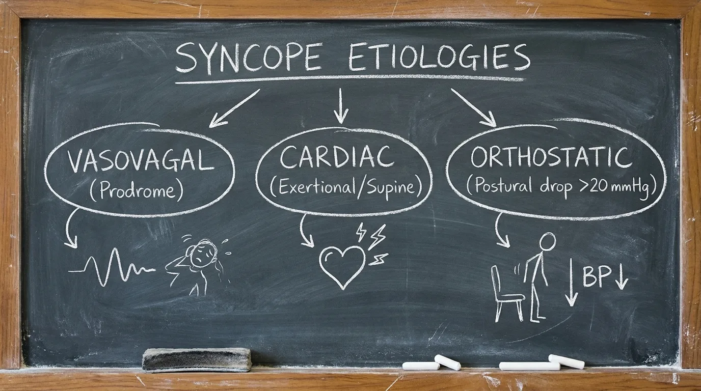
1. Introduction & Etiology Classification
Syncope evaluation requires rapid risk stratification to identify patients at high risk for sudden cardiac death.
---
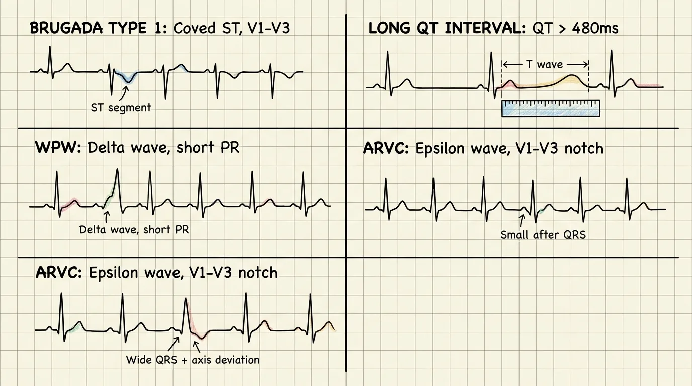
2. Diagnostic Workup & ECG Red Flags
A 12-lead ECG is mandatory in EVERY patient presenting with syncope.
---
3. Cardiac vs Reflex Syncope Differentiation
| Clinical Characteristic | Cardiac Syncope (High Risk) | Vasovagal / Reflex Syncope (Low Risk) |
|---|---|---|
| Trigger / Context | Syncope during exertion, supine position, or sudden loud noise | Prolonged standing, warm environment, venipuncture, pain |
| Prodrome | Absent or sudden brief palpitation (< 5 seconds) | Nausea, diaphoresis, lightheadedness, warmth, tunnel vision |
| History | Known structural heart disease, heart failure, family sudden death | Recurrent episodes since youth, normal cardiac history |
| ECG Findings | Conduction blocks, QTc prolongation, ischemic shifts, Q waves | Completely normal 12-lead ECG |
---
4. San Francisco & Canadian Syncope Risk Scores
Risk stratification rules guide decision-making for hospital admission versus outpatient discharge:
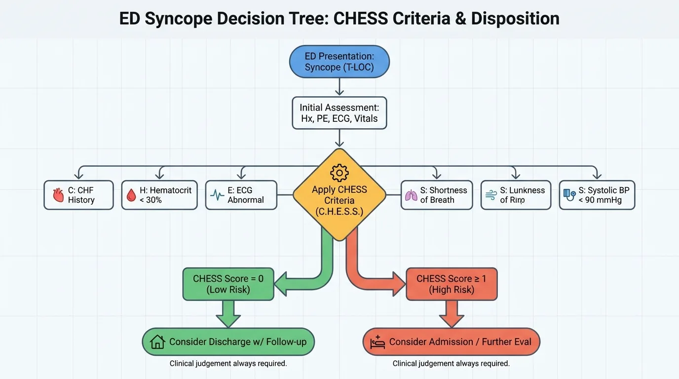
| San Francisco Rule (CHESS Mnemonic) | Clinical Criteria | High-Risk Cutoff |
|---|---|---|
| C - CHF | History of Congestive Heart Failure | Any 1 positive criterion = High Risk (Admit for telemetry) |
| H - Hematocrit | Hematocrit < 30% | High Risk |
| E - ECG | Any non-sinus rhythm or new ECG abnormality | High Risk |
| S - Shortness of breath | History or presentation of acute dyspnea | High Risk |
| S - Systolic BP | Systolic BP < 90 mmHg at presentation | High Risk |
---
5. Structural & Channelopathy Red Flags Table
| Channelopathy / Disease | Pathognomonic ECG Finding | Diagnostic Risk | Immediate Action |
|---|---|---|---|
| Brugada Syndrome | Type 1 coved ST elevation ≥ 2 mm in V1–V2 with T inversion | Lethal polymorphic VT / VF | EP referral + ICD evaluation |
| Long QT Syndrome | QTc > 500 ms (or Bazett formula correction) | Torsades de Pointes risk | Avoid QTc drugs + Beta-blockers |
| ARVC / ARVD | Epsilon wave (notch at end of QRS) in V1–V3 | Right ventricular VT | Avoid competitive sports + ICD |
| Hypertrophic Cardiomyopathy (HCM) | Dagger-like Q waves in lateral leads + LVH voltage | Exertional syncope / Outflow tract obstruction | Echo + Beta-blockers |
---
6. Advanced Diagnostic Equations
▶ Bazett QTc Calculation Formula
Formula: QTc = QT Interval (seconds) / √[RR Interval (seconds)]
- Normal Limits: Males < 450 ms | Females < 460 ms.
- Torsades Risk: QTc > 500 ms represents high risk for lethal ventricular arrhythmias.
---
7. Flagship Worked Clinical Case Study
Worked Case: Syncope Secondary to Unmasked Brugada Syndrome
Case StudyA 34-year-old male with no prior medical history experiences sudden syncope while resting in bed during a febrile viral illness (Temperature 38.8°C).
#### 📊 Patient Lab & ECG Panel
| Diagnostic Parameter | Measured Value | Reference Range | Diagnostic Interpretation |
|---|---|---|---|
| ECG Lead V1–V2 | Coved 2.5 mm STE + T inversion | Isoelectric | Type 1 Brugada Pattern |
| QRS Duration | 104 ms | <120 ms | Normal QRS duration |
| Hs-Troponin | < 0.01 ng/mL | <0.04 ng/mL | Normal (No ischemic necrosis) |
| Echocardiogram | Normal LVEF 62% | >55% | Structurally normal heart |
| Family History | Unexplained sudden death (uncle at 36 yr) | Negative | High-Risk Genetic History |
#### ⚡ Step-by-Step Clinical Decision Flowchart
Step 1: Clinical Context  → Supine syncope during fever + Family history of sudden death = High Cardiac Risk
Step 2: ECG Recognition  → Pathognomonic Type 1 Brugada pattern (Coved STE in V1–V2)
Step 3: Immediate Rx     → Treat fever aggressively with IV Acetaminophen (Fever unmasks sodium channel block!)
Step 4: Admission Action → Admit to Cardiac Telemetry / ICU (CHESS Rule positive for abnormal ECG)
Step 5: EP Management   → Stat Electrophysiology (EP) consult for ICD (Implantable Cardioverter-Defibrillator)
---
8. High-Yield Clinical Pearls & Diagnostic Traps
Clinically Related Case Posters
Sudden Fast Irregular Heartbeat with Breathlessness
Essential Hypertension (15 years, on treatment). Type 2 Diabetes Mellitus (...
Sudden Bluish Discoloration and Crying in a Toddler
Down Syndrome
Sudden Shortness of Breath and Chest Pain
None
Test Your Decision-Making in Syncope Secondary to Brugada Syndrome
Experience this exact clinical scenario in the Bedside Rounds interactive simulator. Order investigations, manage door-to-needle timing, and witness real-time physiological feedback.