Tetralogy of Fallot Clinical Review: Diagnosis & Tet Spell Resuscitation
Master Tetralogy of Fallot pathophysiologic 4 defects, hypercyanotic Tet spell emergency management, boot-shaped heart sign, and surgical timing.
Table of Contents (10 Sections)
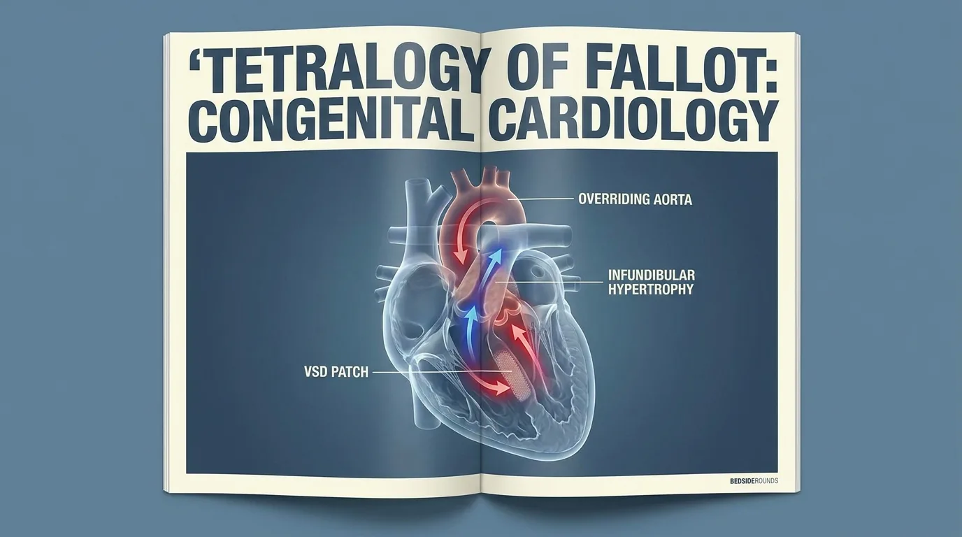
Tetralogy of Fallot Clinical Review: Diagnosis & Tet Spell Resuscitation
3 to 6 months of age.---
📌 Interactive Table of Contents (Index)
- 1. Introduction & 4 Anatomical Defects
- 2. Pathophysiology & Shunt Dynamics
- 3. Hypercyanotic Tet Spell Resuscitation Protocol
- 4. Diagnostic Workup & Boot-Shaped Heart Sign
- 5. Pharmacotherapy & Surgical Repair Timing Table
- 6. Advanced Hemodynamic Equations
- 7. Flagship Worked Clinical Case Study
- 8. High-Yield Clinical Pearls & Diagnostic Traps
---
1. Introduction & 4 Anatomical Defects
Tetralogy of Fallot (TOF) is the most common cyanotic congenital heart defect, accounting for 7–10% of all congenital cardiac malformations.
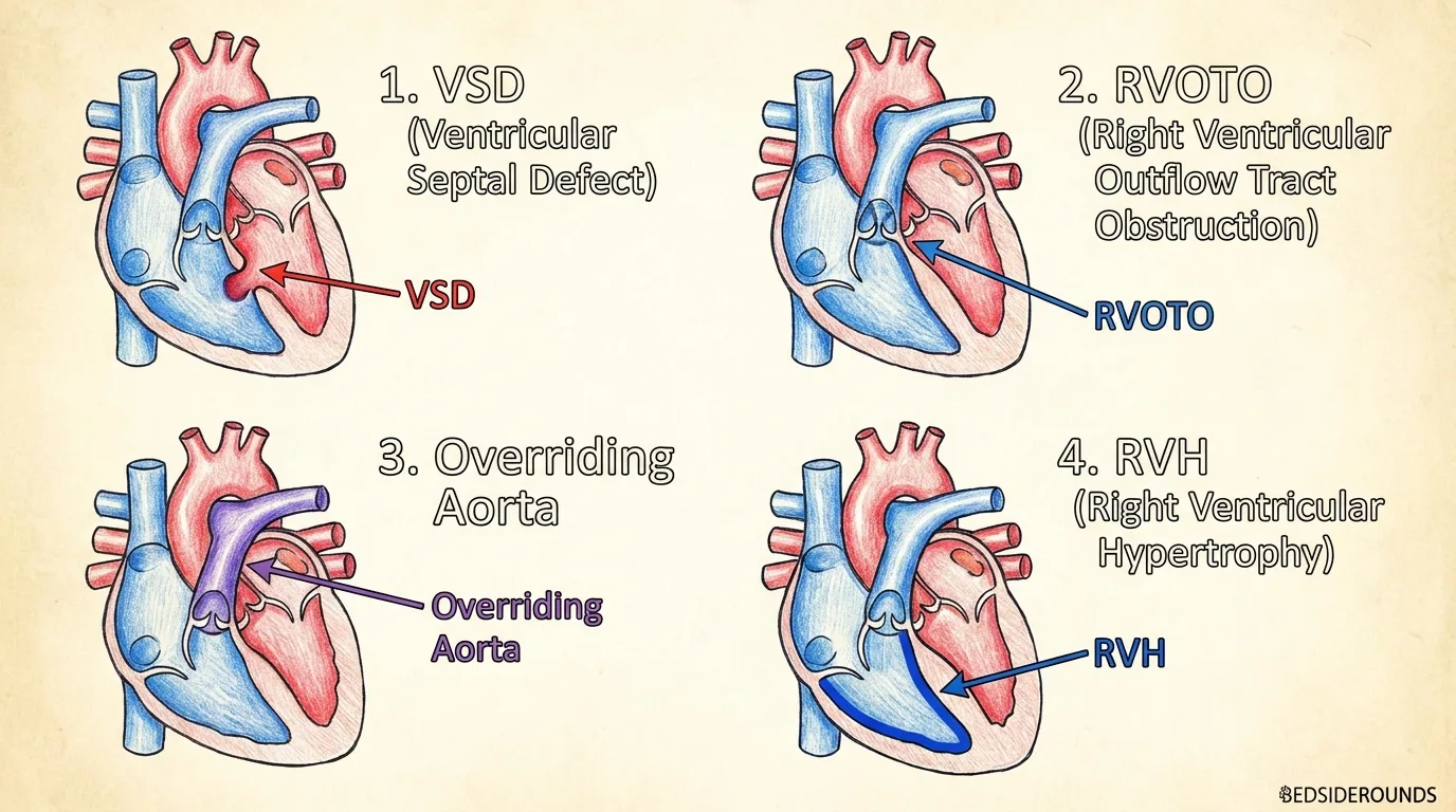
| Anatomical Defect | Pathophysiologic Mechanism | Clinical Manifestation |
|---|---|---|
| 1. RVOT Obstruction | Infundibular stenosis / Subvalvular pulmonary narrowing | Determines degree of cyanosis & right-to-left shunt |
| 2. Malalignment VSD | Large non-restrictive subaortic ventricular defect | Allows equal pressure between LV and RV |
| 3. Overriding Aorta | Aorta positioned directly above VSD (< 50% displacement) | Receives blood from both right and left ventricles |
| 4. Secondary RVH | Right ventricular muscle hypertrophy due to high RVOT pressure | Concentric thickening of right ventricle |
---
2. Pathophysiology & Shunt Dynamics
The direction and magnitude of blood flow across the VSD depends entirely on the ratio of SVR (Systemic Vascular Resistance) to PVR (Pulmonary Vascular Resistance):
- SVR > PVR: Left-to-right shunting (Minimal cyanosis; "Pink Tet").
- PVR > SVR (or Severe RVOT Spasm): Right-to-left shunting (Severe cyanosis; "Blue Tet" / Tet Spell).
---
3. Hypercyanotic Tet Spell Resuscitation Protocol
Follow this emergency resuscitation algorithm for acute hypercyanotic spells:
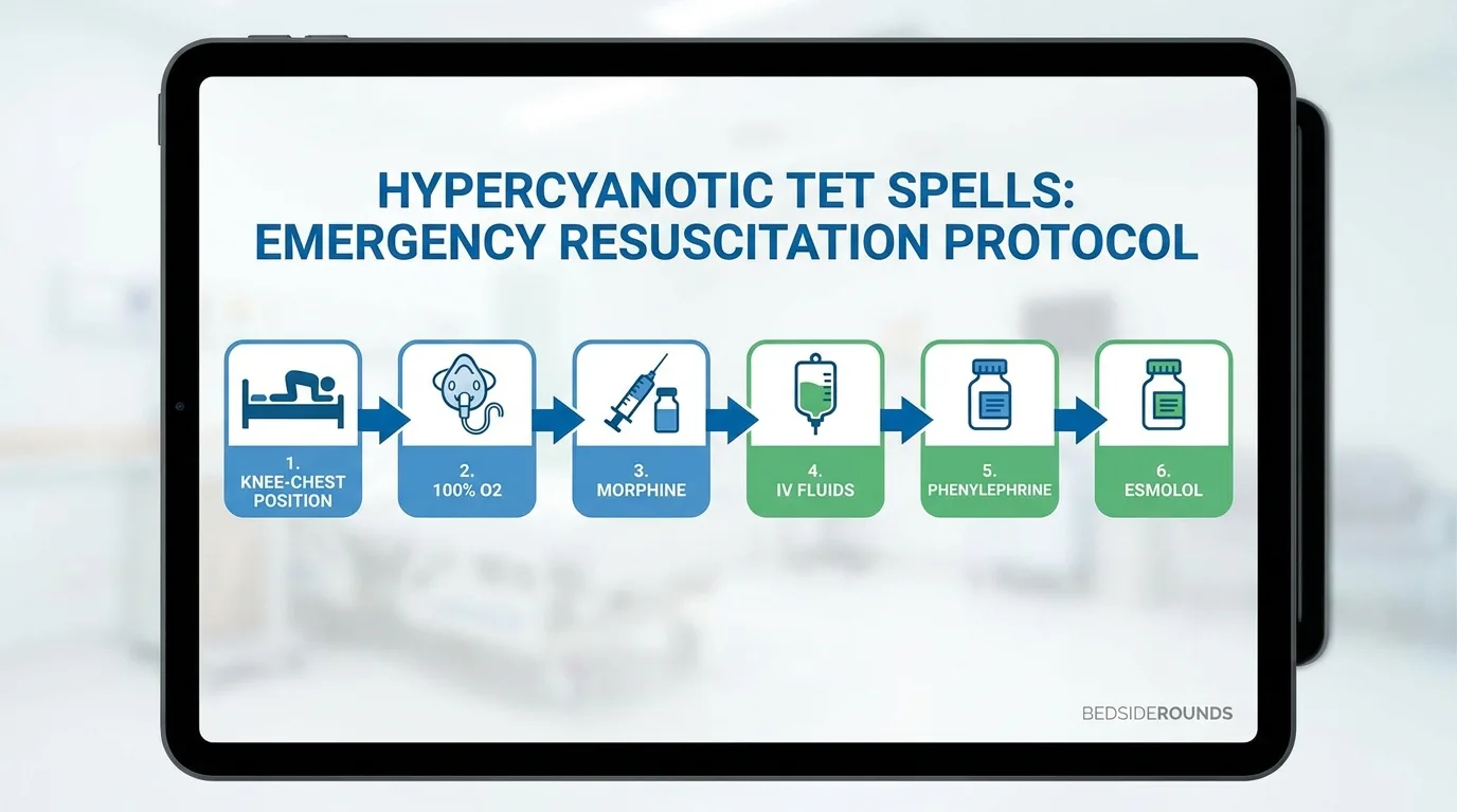
- Step 1: Physical Maneuver (Knee-to-Chest)
- - Flex infant's knees tightly against chest (or squatting in older children). Increases SVR instantly!
- Step 2: Oxygenation & Sedation
- - High-flow 100% O₂ + IV Morphine (
0.1 mg/kg IM/IV) to reduce infundibular spasm and calm agitation. - Step 3: Volume Expansion
- - Administer IV Normal Saline bolus (
10–20 mL/kg) to increase RV preload and pulmonary flow. - Step 4: Pharmacotherapy (Phenylephrine / Propranolol)
- - IV Phenylephrine (
5–20 mcg/kg IV) to raise SVR OR IV Esmolol/Propranolol to relax infundibular muscle.
---
4. Diagnostic Workup & Boot-Shaped Heart Sign
- Chest X-Ray: Classic "Boot-shaped heart" (Coeur en sabot) due to RV hypertrophy elevating the apex + dark oligemic lung fields (decreased pulmonary vascular markings).
- 12-Lead ECG: Right axis deviation (+120° to +180°) + Right Ventricular Hypertrophy (tall R wave in V1, deep S wave in V6).
- Echocardiogram: Definitive gold standard to visualize VSD, RVOT gradient, and coronary anatomy.
---
5. Pharmacotherapy & Surgical Repair Timing Table
| Phase / Intervention | Indication / Dosing | Primary Objective |
|---|---|---|
| PGE1 (Alprostadil) | 0.05–0.1 mcg/kg/min IV infusion | Keeps Patent Ductus Arteriosus (PDA) open in duct-dependent newborns |
| Oral Propranolol | 0.5–1.0 mg/kg/dose PO tid | Prevents recurrent infundibular spasms while awaiting elective surgery |
| Complete Surgical Repair | Elective at 3–6 months of age | VSD patch closure + RVOT transannular patch reconstruction |
| Palliative BT Shunt | Modified Blalock-Taussig-Thomas shunt | Temporary systemic-to-pulmonary shunt in premature / low birth weight infants |
---
6. Advanced Hemodynamic Equations
▶ Qp / Qs Ratio (Pulmonary-to-Systemic Flow Ratio)
Formula: Qp / Qs = (SaO₂ - SvO₂) / (SpvO₂ - SpaO₂)
- Normal Ratio:
Qp / Qs = 1.0 - Cyanotic Right-to-Left Shunt (TOF):
Qp / Qs < 1.0(Pulmonary blood flow is severely reduced).
---
7. Flagship Worked Clinical Case Study
Worked Case: Hypercyanotic Tet Spell in a 7-Month-Old Infant
Case StudyA 7-month-old infant with uncorrected Tetralogy of Fallot presents to the ED during a crying episode with severe cyanosis, hyperpnea, and SpO₂ of 54%.
#### 📊 Patient Lab & Pediatric Panel
| Diagnostic Parameter | Measured Value | Reference Range | Diagnostic Interpretation |
|---|---|---|---|
| SpO₂ (Room Air) | 54% | 95% – 100% | Severe Hypoxemic Cyanosis |
| Heart / Resp Rate | 178 bpm / 62 bpm | 100–140 / 25–40 | Tachypnea & Sympathetic Surge |
| Chest X-Ray | Boot-shaped Heart | Normal | Pathognomonic RV Apex Elevation |
| Capillary Blood Gas | pH 7.19 / PaO₂ 24 | pH 7.35–7.45 | Severe Metabolic Lactic Acidosis |
| Cardiac Auscultation | Loss of Systolic Murmur | Grade 3/6 murmur | Complete RVOT Obstruction Spasm |
#### ⚡ Step-by-Step Clinical Decision Flowchart
Step 1: Immediate Maneuver → Place infant in tight Knee-to-Chest position (Increases SVR to force flow into lungs)
Step 2: Oxygen & Calming  → High-flow 100% O₂ + IV Morphine 0.1 mg/kg IV (Calms agitation & relaxes RVOT)
Step 3: Volume Resus     → Administer IV Normal Saline 20 mL/kg rapid bolus (Increases RV filling pressure)
Step 4: Vasopressor Push  → Give IV Phenylephrine 10 mcg/kg IV (Elevates SVR above PVR → Shunt reverses!)
Step 5: Outcome           → SpO₂ rises to 88%, Systolic murmur returns (RVOT spasm relieved!), stat Echo ordered
---
8. High-Yield Clinical Pearls & Diagnostic Traps
Clinically Related Case Posters
Sudden Bluish Discoloration and Crying in a Toddler
Down Syndrome
Rapid Breathing and Feeding Difficulty in an Infant After a Cold
Previously healthy, full-term infant (39 weeks, 3.2 kg birth weight). No NI...
High Fever and Stiff Neck in a 4-Year-Old
Previously healthy child with no chronic illnesses. All vaccinations up to ...
Test Your Decision-Making in Hypercyanotic Tet Spell in an Infant with Uncorrected TOF
Experience this exact clinical scenario in the Bedside Rounds interactive simulator. Order investigations, manage door-to-needle timing, and witness real-time physiological feedback.